Schwannoma
A Schwannoma (the plural is actually Schwannomata but many people say Schwannomas) is a lump in a nerve .
The word ‘tumour’ worries people but it is a medical word that just means ‘lump’.
Schwannomata are ‘tumours’ (they are lumps or growths) but they are NOT cancer.
These lumps grow from cells called Schwann cells (if you are interested they were named after the man who first saw them down a microscope and wrote about them a man called Theodor Schwann in 19th Centuary Bonn, Germany).
Schwannomata grow within nerves. They come from one part of the nerve and grow inside the outlining of the nerve making a lump on the nerve.
How is a Schwannoma diagnosed?
The commonest presentation is an incidental lump, found by the individual (when washing for example, or as it is noticed that press on a particular area of the body is uncomfortable or it creates tingling further down the limb).
All lumps need to be examined by a doctor as some might be cancerous.
The Schwannoma lump should under go a full examination physically and then an MRI scan. An MRI (a scan where you lie in a big donught shaped scanner which makes funny squeaky noises as magnets are used to take pictures of your body) provides a pictures which allows in most cases a diagnosis to made, along with the findings from the patients story of their symptoms and the examination the doctor did. The diagnosis of Schwannoma from examination and MRI scanning is very close to being right all the time in most cases -around 98 or 98%. However it cannot be said for certain 100% that the lump is not cancer; The only way to be completely sure is to take the lump out with surgery.
Surgery has risks and so should only be undertaken for the following reasons:
1. If the lump cannot be said to be non-cancerous with great enough certainty.
2. If the symptoms from the lump are intrusive
3. If the are other concerns about the lump.
4. If the patient after understanding the risks and benefits wishes to have the lump removed.
So if the symptoms are not too bad and the examination and scan are typical for a Schwannoma, then it is certainly fine to not have surgery.
Biopsy?
Some people suggest needle biopsy of these lumps (taking a bit our using a needle through the skin and into the lump) but I am strongly against this as it damages the normal parts of the nerve, can cause severe pain not just during the biopsy but for a long time afterwards.
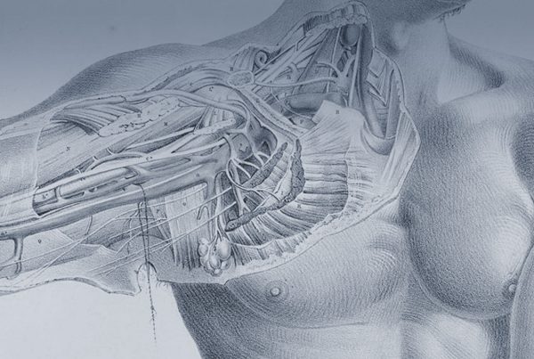
I recommend open excisional biopsy of the tumor (as is illustrated above) to remove the lump- so it is no longer a problem and we can find out what kind of lump is was (confirm it was a schwannoma).
What does surgery mean to me?
The aims of surgery are to remove the lump to
1. Gain a diagnosis
2. Improve and hopefully remove the symptoms
The risks are:
1. Infection
2. Scar tenderness or pain
3. Damage to the nerve’s function
4. Pain
5. Stiffness
6. Bleeding
7. Lack of improvement in symptoms
8. Bloodclots (DVT, PE)
The implications:
To understand the issues around risk and benefit and elect with all the information available (regarding surgery and all the options including non- operative management) decide to proceed to surgery
To undergo surgery to remove the tumour it will be necessary to undergo the following:
- pre-assessment protocols (to ensure best health and to exclude any carriage of MRSA bacteria and SRA-CoV2 COVID virus).
- To be admitted to hospital on the morning of surgery and be nil by mouth from the night before (clear water or black tea without lemon or milk is ok unto 2 hours before operation but is best avoided for 4 hrs prior).
- To sign the consent form (re-afirming the consent in a written matter) and have a mark placed to identify the correct surgical site.
- Surgery often lasts around 2hrs in total.
- After waking from surgery it is often possible to return home the same night : This is not possible sometimes if you are not completely comfortable following surgery or if the operation was later in the day and discharge would be very late and in which case you will stay over night following surgery an be able to return home the next day.
- There will be dressings over the wound which will stay in place until two weeks after surgery.
- Depending on your work Mr Quick would suggest that you take two weeks off work post operatively to ensure the best outcome, some people can return to light desk based work in some cases.
Follow up
It is Mr Quick’s standard practice that following surgery you are reviewed by Mr Quick directly after surgery, then if you are still in hospital the next day.
Then a wound check at two weeks after the operation
The a full clinic assessment at six weeks following surgery
Then at three months following surgery